• 450-967-0500
  • [email protected]
  • Français
  • Home
  • Treatments
        • SPORTS MEDICINE
          • Sports Medicine
          • Brain Concussion
          • Ultrasound Infiltration
          • Treatment of osteoarthritis
        • PHYSIOTHERAPY
          • Physiotherapy
          • Pelvic and Perineal Rehabilitation
        • CHIROPRACTIC
          • Chiropractic
          • Functional Range Release Technique
          • Active Release Techniques
          • Graston
          • Fascial Manipulation
          • Fascial Distortion Model
          • Cupping
          • Kinesio Taping / Rocktape
          • Neurocryotherapy
          • Low intensity laser
          • Shockwave Therapy
          • Neurorehabilitation in functional neurology
        • OTHERS
          • Massage therapy
  • Conditions
    • Back Pain
    • Headaches and Neck Pain
    • Legs Pain
    • Arms Pain
    • Feet Pain
  • Our team
  • Clinic
  • Contact Us
Search
Close

Arms Pain

Several conditions may be related to arm pain. Here are some examples of the conditions we are dealing with. Of course, this list is not exhaustive, so for more details, contact us.
  • Medial Epicondylitis (Golfer elbow)
  • Bursitis / Tendinitis of the shoulder
  • Lateral epicondylitis (Tennis elbow)
  • Bursitis
  • Trigger finger
  • Carpal tunnel syndrome
  • Adhesive Capsulitis (Frozen Shoulder)
  • Osteoarthritis
01

Medial Epicondylitis (Golfer elbow)

Description
  • Medial epicondylitis also occurs during repetitive movements involving the wrist, whether at work or during daily activities.
  • It can also occur in golfers, whether amateur or professional.
  • Following overwork, inflammation occurs where the muscles and tendons attach to the elbow. The muscles and tendons involved are those that allow flexion of the wrist and fingers.
  • Without proper treatment, this condition can develop into a chronic and recurring problem. The presence of degeneration or partial tearing of the tendon is possible.
Presentation
  • The pain is on the inside of the elbow.
  • It will be felt when the inflamed muscle or tendon is used. At first, it will be during specific activities and then manifest in simple daily activities such as lifting a glass or turning a doorknob. A weakness in these activities may also be noted.
Causes
  • Repetitive stress and chronic injuries involving the musculature and tendons of the forearm
  • Weakness and imbalance of the muscles
  • Abuse of exercises (syndrome “too much effort, too early”)
  • Unsuitable equipment
  • Bad technique
Treatments
  • Modification of activities that are associated with the condition
  • Reduction of inflammation with ice (20 minutes / 2 hours) and complementary therapies including neuro-mediotherapy
  • Joint manipulations and soft tissue work (Active Release Techniques and Graston) to correct the biomechanics of the elbow and reduce scar tissue
  • Stretching exercises
  • Exercises to strengthen and stabilize the elbow
  • Medications (anti-inflammatories) as needed
  • Orthopedic appliance and kinesio taping to reduce the pressure on the damaged tendon.
  • Injections of cortisone as needed
  • Very rare surgical procedures
  • Plasma rich in platelets
Prognosis
  • Acute condition, for which symptoms have been present for less than 4 weeks, is treated in 2 to 6 weeks.
  • Chronic condition, for which symptoms have been present for more than 8 weeks, may require 3 to 6 months.

Bursitis / Tendinitis of the shoulder

Description

Tendinitis or bursitis of the shoulder is one of the most common injuries among those who participate in sports involving arm extension above the head (eg swimming and baseball). It is an inflammation of the tendons or bursae that pass through a narrow channel, even more restricted when the arm is raised.

Presentation
  • Pain is usually felt in the upper part of the shoulder or lower in the muscle of the shoulder.
  • Occurs when the arm is raised above the head or during certain twisting movements.
  • In extreme cases, the pain may be constant and may prevent the person from sleeping.
Causes
  • Abuse of exercises (syndrome “too much effort, too soon”) especially the arm above the head
  • Weak and unbalanced muscles accentuating pressure on the tendons and thus inflammation
  • Bad technique in a sport
  • Hard training
  • Loose shoulder joint for genetic or injury reason
  • Bad posture
  • Dysfunction of the scapula
  • Injury anterior to the shoulder
Treatments
  • Reduction of inflammation with ice and electrotherapy and neurocryotherapy
  • Range of motion to prevent the shoulder from stiffening
  • Joint manipulations and soft tissue work (Active Release Techniques and Graston) to correct the biomechanics of the shoulder and reduce scar tissue
  • Exercises to strengthen and stabilize the shoulder
  • Modification of activities
  • kinesio taping
  • Medications (anti-inflammatories) as needed
  • Injections of cortisone as needed
  • Very rare surgical procedures
  • Plasma rich in platelets

Lateral epicondylitis (Tennis elbow)

Description
  • Lateral epicondylitis is the most common elbow injury. It affects many racquet sports participants, whether amateur or professional.
  • This injury also occurs during repetitive movements involving the wrist, whether at work or during daily activities.
  • Following overwork, inflammation occurs where the muscles and tendons attach to the elbow. The muscles and tendons involved are those that allow extension of the wrist and fingers.
  • Without proper treatment, this condition can develop into a chronic and recurring problem. The presence of degeneration or partial tearing of the tendon is possible.
Presentation
  • The pain is on the outside of the elbow.
  • It will be felt when the inflamed muscle or tendon is used. At first, it will be during specific activities and then manifest in simple daily activities such as lifting a glass or turning a doorknob. A weakness in these activities may also be noted.
Causes
  • Repetitive stress and chronic injuries involving the musculature and tendons of the forearm
  • Weakness and imbalance of the muscles
  • Abuse of exercise (syndrome “too much effort, too early”)
  • Unsuitable equipment
  • Bad technique
Treatments
  • Modification of activities that are associated with the condition
  • Reduction of inflammation with ice (20 minutes / 2 hours) and complementary therapies including neuro-mediotherapy
  • Joint manipulations and soft tissue work (Active Release Techniques and Graston) to correct the biomechanics of the elbow and reduce scar tissue
  • Stretching exercises
  • Exercises to strengthen and stabilize the elbow
  • Medications (anti-inflammatories) as needed
  • Orthopedic appliance and kinesio taping to reduce the pressure on the damaged tendon.
  • Injections of cortisone as needed
  • Very rare surgical procedures
  • Plasma rich in platelets
Prognosis
  • Acute condition, for which symptoms have been present for less than 4 weeks, is treated in 2 to 6 weeks.
  • Chronic condition, for which symptoms have been present for more than 8 weeks, may require 3 to 6 months.

Book an appointment now

If pain persists and you need help, our multidisciplinary team will be able to get you back on track quickly.

Contact Us
Our team
04

Bursitis

Description
  • A purse is similar to a soft, fluid-filled bag that cushions bones and soft tissues such as tendons and skin.
  • Bursitis is an inflammation of the purse.
  • Typically affected bursaries are at the shoulders, elbows, knees, pelvis, hips, heels and Achilles tendons.
  • Common problem but not serious.
Presentation
  • Pain, tension and movement limitation in the affected area, occasionally associated with pain radiating to adjacent areas.
  • Occasionally, severe pain is felt when moving the affected area.
  • The swollen pouch may be painless.
  • Possibility of fever if there is an infection.
Treatments
  • Rest and modification of the activities that are associated with the involved articulation
  • Reduction of inflammation with ice (20 minutes / 2 hours), electrotherapy and neuro-therapy
  • Joint manipulations and soft tissue work (Active Release Techniques and Graston) to correct the biomechanics of the joint and reduce scar tissue
  • kinesio taping
  • Medications (anti-inflammatories) as needed
  • Stretching exercises
  • Exercises to strengthen and stabilize the joint
  • If necessary, remove the liquid from the purse with or without cortisone injections
  • Very rare surgical procedures involving removing the purse involved
Prognosis
  • Symptoms usually decrease between 7 and 14 days.
Causes
  • Direct injury at a joint or stock exchange
  • Overload or unusual exercise involving a joint, especially running on an incline or downhill (knees)
  • Prolonged pressure on the purse (leaning on the elbows or kneeling)
  • Deposition of calcium in the tendons of the shoulder with tendon degeneration
  • Acute or chronic infection
  • Arthritis
  • Drop

Trigger finger

Description

The flexor muscles of the fingers are located in the forearm and tendons attach to the small bones of the fingers and thumb. When we flex or straighten the fingers, the flexor tendons slide into a small tunnel, called the tendon sheath, which keeps the tendon in place near the bones. The flexor tendon may progressively become irritated under this tendinous sheath. This will gradually result in a thickening of the tendon and the probable appearance of nodules causing a much more difficult passage of the tendon in the tunnel. In the presence of finger (or thumb) trigger, the tendon will be temporarily blocked during passage through the tunnel (tendon sheath) during the flexion / extension of the finger.

Presentation
  • Presence of a sensitive hump in the palm of the hand.
  • Swelling.
  • Sudden blocking and unblocking sensation during flexion / extension of the finger.
  • Pain during flexion / extension of the affected finger.
Causes
  • Unknown cause.
  • More common in women.
  • More common among 40 to 60 year olds.
  • People with diabetes or rheumatoid arthritis are at higher risk.
Treatments
  • If very light, rest can help.
  • Treatment of Active Release Techniques (ART) or Graston to break down muscle and tendon adhesions.
  • Use of neuro-therapy, ice, or NSAIDs to reduce inflammation or pain.
  • Stretching exercises and modifying the activities associated with the condition.
  • Injection of cortisone in cases refractory to conservative treatment.
  • Surgery in cases refractory to cortisone.
Prognosis
  • In mild to moderate cases, improvement is often noted after 4-6 weeks of conservative treatment.
  • In cases where surgery is needed, most cases will return to normal movement immediately after surgery, but a period of 6 months may be necessary for swelling and stiffness to completely disappear.
05

Carpal tunnel syndrome

Description

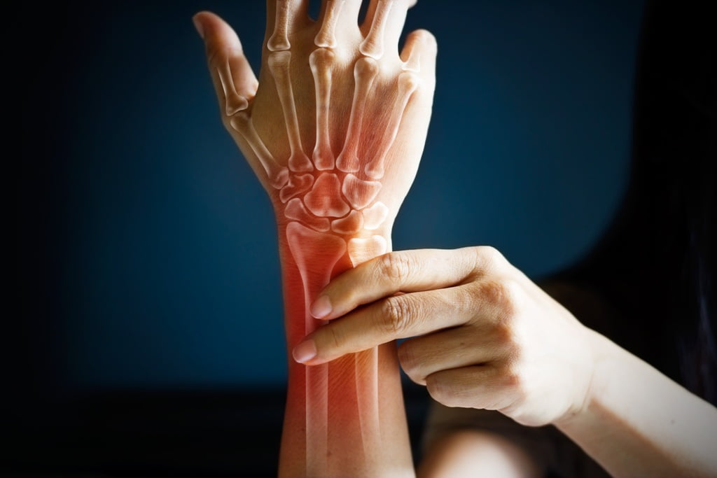
The carpal tunnel is a small narrow tunnel located at the wrist. Some carpal bones and the transverse carpal ligament define the tunnel. The median nerve travels from the forearm to the hand as it passes through this tunnel. This nerve controls the sensation of the palmar surface of the first 3 fingers and the muscles of the base of the thumb. The tendons of the finger flexors also pass through this tunnel. When the tissues around the tendons of the flexors become irritated and swollen, this can cause pressure on the median nerve, causing the carpal tunnel syndrome. The median nerve may also be irritated or compressed by certain muscles during its passage in the arm, particularly in the pronatoric round muscle.

Presentation
  • Numbness, pecking and pain in the hand (median nerve territory).
  • Sensation of electric shock at the first 3 fingers (from the thumb).
  • A strange sensation that can radiate into the arm and shoulder.
  • Symptoms are very common at night.
Causes
  • The carpal tunnel can be anatomically smaller in a hereditary way.
  • Over-use of the hand and forearm causing soft tissue adhesions (muscles, ligaments) compressing the median nerve.
  • Hormonal changes (eg pregnancy)
  • Certain medical conditions, such as diabetes, rheumatoid arthritis and some thyroid disorders.
Treatments
  • Following the diagnosis, changes to the activities associated with the condition.
  • Treatment of Active Release Techniques (ART) or Graston to break up adhesions in structures that can compress the median nerve.
  • Treatment of neurocryotherapy to reduce inflammation and edema in the carpal tunnel.
  • Stretching and rehabilitation exercises.
  • Use a night orthosis to keep the wrist in a neutral position.
  • Use as needed of NSAIDs (anti-inflammatory) and cortisone injection.
  • Decompression surgery may be necessary in some cases refractory to conservative treatment.
Prognosis
  • In many cases, the conservative approach will result in a cure of the condition within a few weeks or months.
  • In severe cases, even after surgery, symptoms may reappear and a second surgery may be necessary.
07

Adhesive Capsulite

Presentation

Adhesive capsulitis is a pain that is often very intense in the shoulder accompanied by marked loss of movement. It is divided into three phases that can be spread over more than 2 years. The first being the painful phase, the second being the “frozen” phase and the last being the recovery phase.

Causes
  • Idiopathic (unknown cause).
  • Trauma (tearing of the rotator cuff, for example).
  • Surgery (complication encountered following shoulder surgery).
Treatments
  • The intervention of choice is distensive arthrography, where a quantity of liquid combined with cortisone is injected into the glenohumeral joint.
  • Studies show that combining the previous intervention with manual therapies was more effective than injection alone, hence the importance of post-injection follow-up.
  • Soft tissue techniques (Active Release Techniques and Graston).
  • Passive joint mobilization of the joint.
  • Electrotherapy and ultrasound can also be used to reduce pain, as can applying ice.
  • Rehabilitation exercises should be integrated as soon as possible, where a progressive mobilization of the articulation will be put forward.
Prognosis
  • Without treatment, the condition will resolve itself over a period that may last more than two years.
  • With treatment, it is possible to reduce the recovery time and prevent long-term complications of capsulitis, such as loss of range of motion or prolonged disability.

Osteoarthritis

Description

Osteoarthritis is a degenerative phenomenon attacking the cartilaginous aspect of the joints. Although this degeneration is only the fact of age, it can cause pain in some.

Osteoarthritis is a very common phenomenon in the population, especially in the over 50s. It can still start between 20 and 30 years. Men and women are affected in equal proportions.

Presentation
  • Joint stiffness
  • Decreased movements
  • Decreased flexibility
  • Noise in the joints
Favorite sites
  • Spine
  • Knees and hips (body weight)
Treatments
  • Joint manipulations and soft tissue work (Active Release Techniques and Graston) to improve mobility and reduce scar tissue
  • kinesio taping.
  • Stretching exercises
  • Strengthening exercises
  • Heat (morning stiffness)
  • Changing activities that are associated with pain
  • Medications (anti-inflammatories) as needed
  • Injections of cortisone as needed
  • Very rare surgical procedures
01

Any Questions?

Our multidisciplinary sport medecine team will be happy to help you. Contact us for all your questions or to book an appointment.

  • 450-967-0500
Contact Us
  • Clinic
  • Treatments & Conditions
  • Our Team
  • Contact Us
Menu
  • Clinic
  • Treatments & Conditions
  • Our Team
  • Contact Us
  • Français
© 2025 Clinique de médecine sportive de l’avenir – Développement web par Aponia – Confidentiality Policy